“Wait—Here?!” Discovering the Bird Heart’s Surprising Pump Power Through Real Dissection
This is Ken Kuwako, Science Trainer. Every day is an experiment.
How do birds, soaring high above the clouds, manage to generate the immense energy needed for such vigorous flight? The secret lies hidden within their small bodies: a “super-efficient engine.” This engine is the heart, a marvel of engineering crafted as intricately as, or perhaps even more so than, our own.
Today, I bring you a report on the dissection of a bird heart (chicken heart, or ‘hatsu’ in Japanese) that my students and I conducted during a special 75-minute class session. Through examining this small organ, we explored the mystery of how life efficiently pumps energy throughout the body.
Prepare to be amazed by the exquisite engineering of life—details you can only truly grasp by touching the real thing, not just reading a textbook.
Visualize the Structure First
Before starting the dissection, it is crucial to have a clear understanding of the heart’s basic mechanics. Tracking the history of animal evolution reveals that while fish have a “1 atrium, 1 ventricle” structure, and amphibians have “2 atria, 1 ventricle,” birds and mammals possess the most advanced structure: 2 atria and 2 ventricles.
This design allows for the complete separation of oxygen-rich and oxygen-poor blood. This crucial mechanism supports the high metabolism birds need to maintain high body temperatures and power strenuous flight.
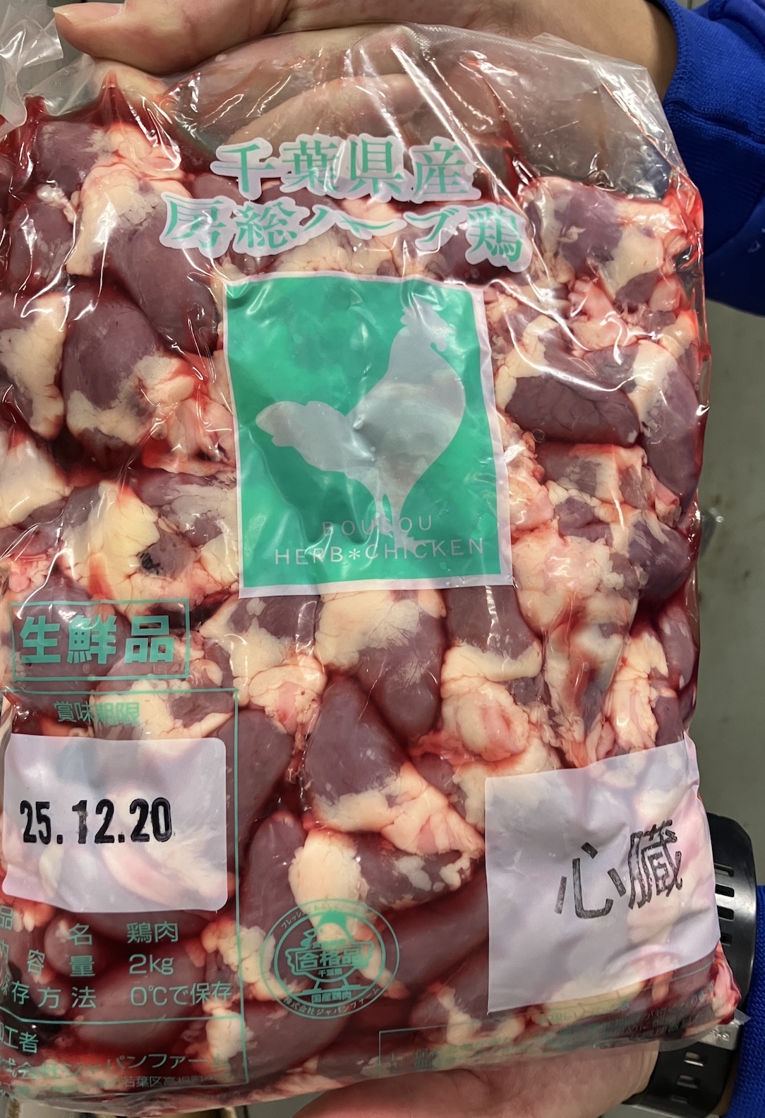
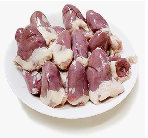
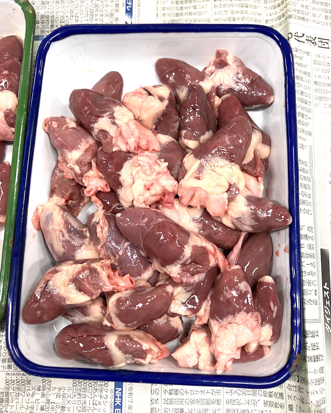
For this class, we used chicken hearts, readily available at most grocery stores. A 2kg pack contained about 130 to 140 hearts, providing an optimal amount for every student in the class to closely observe their own specimen.
To further enhance their mental visualization of the working heart, students watched this instructional video before the dissection.
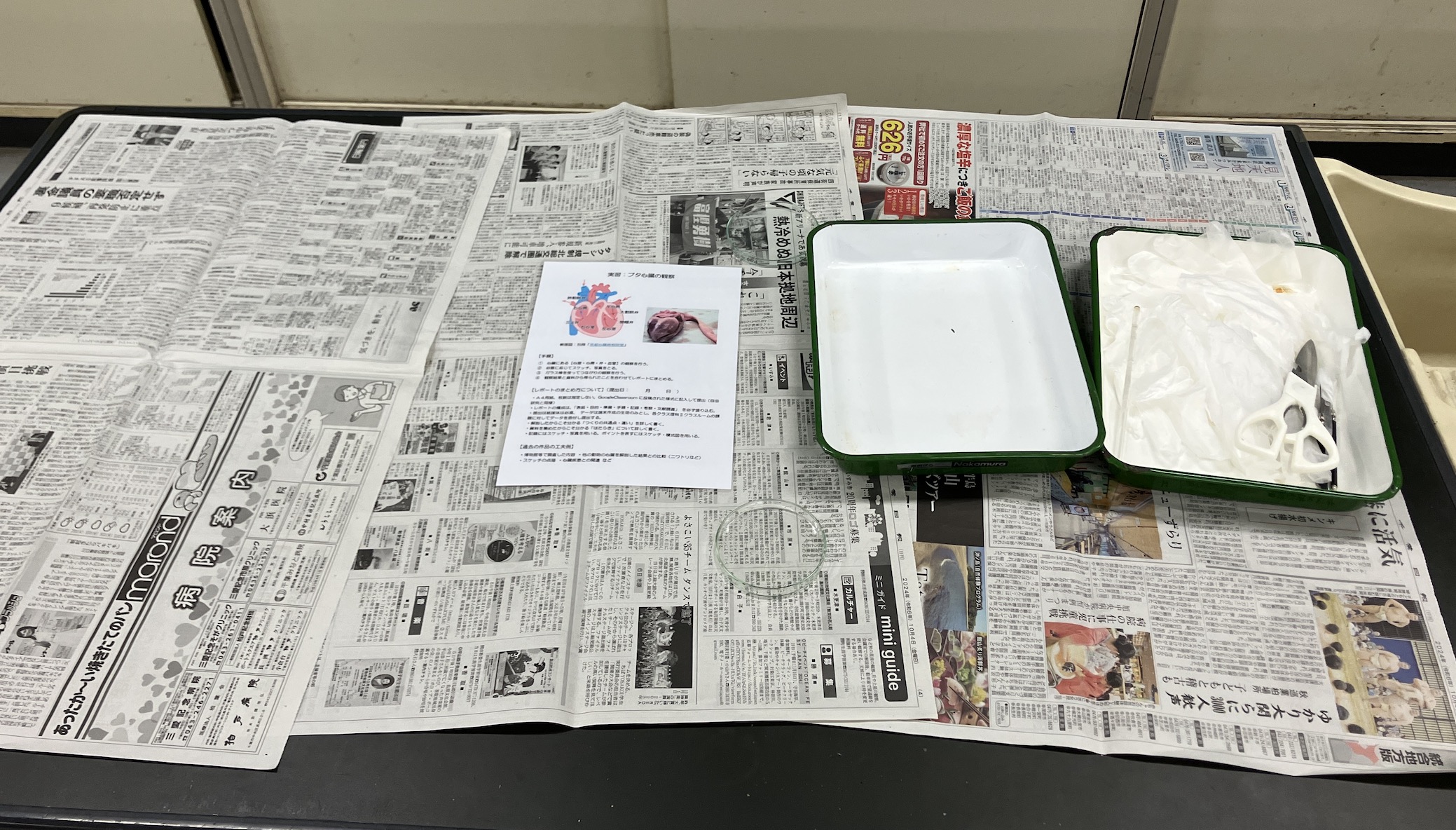
The Kitchen Shears Challenge: Exploring the Heart’s Interior
When we think of dissection, a surgical scalpel often comes to mind. However, for middle school classes, kitchen shears prove to be an exceptionally effective and safe tool. By carefully maneuvering the tips of the shears, we can safely open up the small heart without damaging its delicate internal structures.
Dissection Procedure
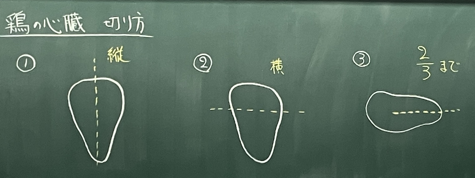
1. The Longitudinal Cut: Halving the Heart! First, we cut the heart in half vertically using the shears to view the cross-section. This is the initial step to confirm that the heart is not just a muscle mass, but contains distinct internal chambers.

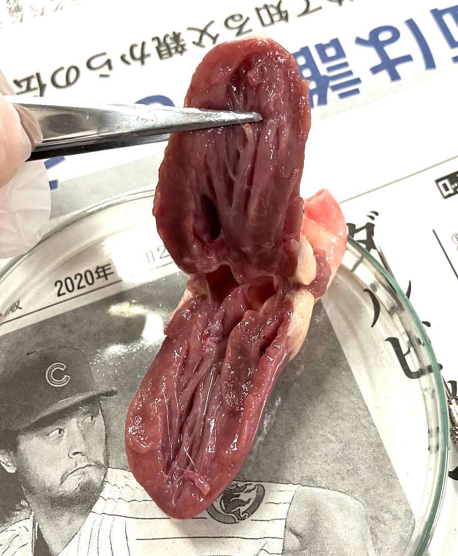
2. The Lateral Incision: Opening it Up To get a more detailed look at the structure, we made a lateral cut, opening the heart up about two-thirds of the way. This technique allowed a panoramic view of how the left ventricle, right ventricle, and the atria are arranged and interconnected.
A Striking Discovery: The Small Right Ventricle and Mighty Left Ventricle
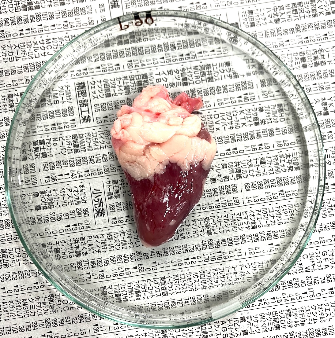
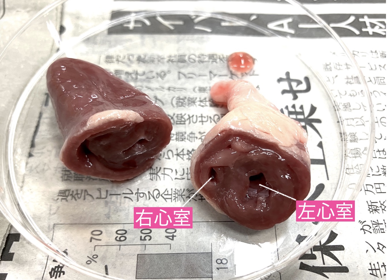
The students were audibly surprised by what they saw when they examined the cross-section. The bird heart is tiny, making the atria a bit challenging to distinguish, but everyone’s attention was drawn to the dramatic difference in size of the ventricles.
Compared to the left ventricle, the bird’s right ventricle is surprisingly thin and small. Some students even questioned, “Is this all there is?” In stark contrast, the wall of the left ventricle, which pumps blood throughout the entire body, was thick, strong, and visibly a solid mass of muscle.
The right ventricle only needs to push blood to the nearby lungs, but the left ventricle must push blood against gravity to every single part of the bird’s body. This vast difference in muscle thickness is the ultimate evidence of life’s strategic design, prioritizing efficiency and power.
Tracing the Blood’s Pathway
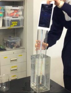
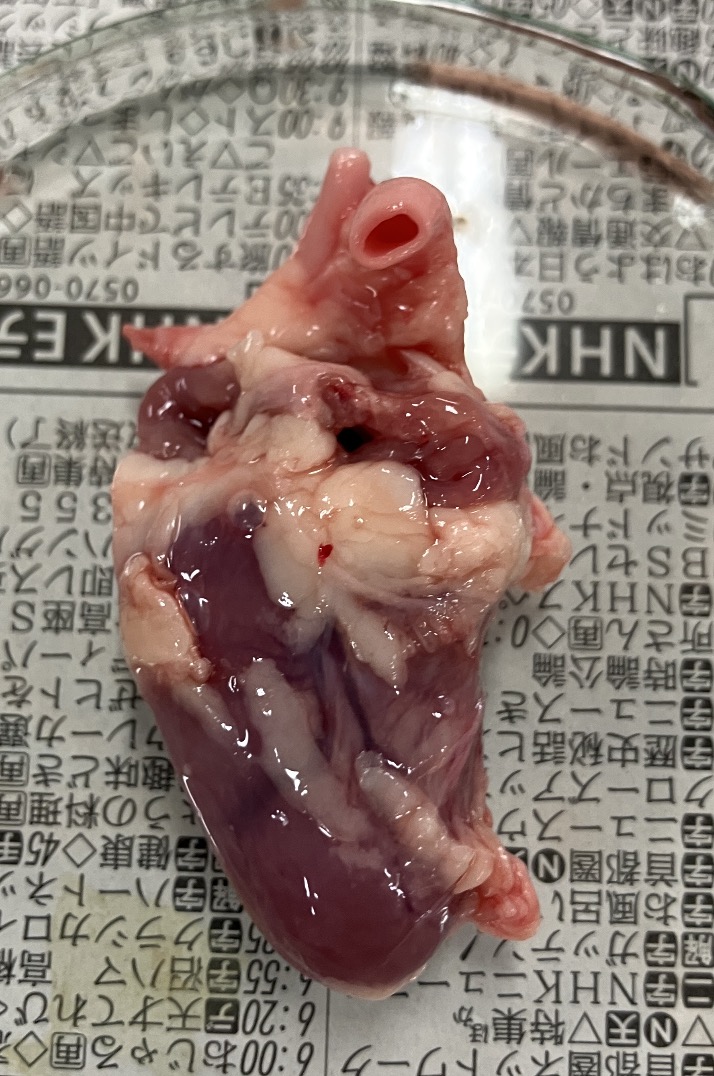
Finally, we conducted a simple experiment to confirm the direction of blood flow. We gently inserted a toothpick into the left ventricle and carefully pushed it forward.
And just like that, the tip of the toothpick smoothly emerged from the aorta!
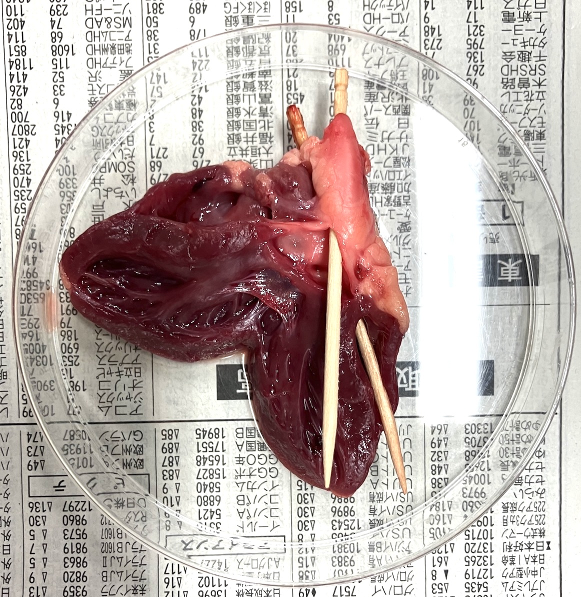
The toothpick closest to the camera is in the aorta, the one further back is in the vena cava.
In that moment, the heart transformed from a collection of parts into a single, perfectly engineered pump system. The sheer reality of the specimen offers an overwhelming power and beauty that diagrams cannot capture.
The small chicken heart taught us the profound value of the organ that tirelessly works every second to sustain our lives. I strongly encourage experiencing this “engineering of life” in your own classroom.
Inquiries and Requests
Discover more about the wonders of science! I compile simple, fun science experiments you can do at home and share helpful tips. Please feel free to search around! ・My science notes have been published as a book. Details can be found here. ・About the administrator, Ken Kuwako, click here. ・For various requests (writing, lectures, science classes, TV supervision, appearances, etc.), click here. ・Article updates are delivered on X!
![]() The Science Content Channel is streaming experiment videos!
The Science Content Channel is streaming experiment videos!
6月のイチオシ実験!
レモンやオレンジで風船を割ろう!インパクトが抜群のリモネン風船の実験
テレビ番組監修・イベント等のお知らせ
- 6月3日(水)20:30〜 「
バカリズムのちょっとバカりハカってみた!」(テレビ東京)を科学監修・出演します。テーマは「 そばの出前は何人前まで運べるのか、限界を測ってみた」です。 - 6月4日(木) 7:00〜 「THE突破ファイル」(日本テレビ)について科学監修しました。
- 6月14日(日) 千葉大学インスタレーション「探究」にて講師を務めます
- 6月26日(金) 公開研究会「脱作業化!デジタル化と段階的指導で実現する オームの法則の探究」
- 6月28日(日) ダビンチマスターズ@昭和女子
- 7月18日(土) 教員向け実験講習会「ナリカカサイエンスアカデミー」の講師をします。お会いしましょう。
書籍のお知らせ
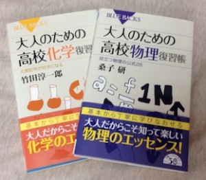
- 『大人のための高校物理復習帳』(講談社)…一般向けに日常の物理について公式を元に紐解きました。特設サイトでは実験を多数紹介しています。※増刷がかかり6刷となりました(2026/02/01)
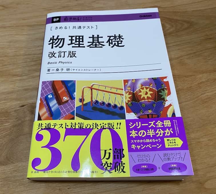
- 『きめる!共通テスト 物理基礎 改訂版』(学研)… 高校物理の参考書です。イラストを多くしてイメージが持てるように描きました。授業についていけない、物理が苦手、そんな生徒におすすめです。特設サイトはこちら。
各種SNS(更新情報をお届け!)
X(Twitter)/instagram/Facebook(日本語)
Explore
- 楽しい実験…お子さんと一緒に夢中になれるイチオシの科学実験を多数紹介しています。また、高校物理の理解を深めるための動画教材も用意しました。
- 理科の教材… 理科教師をバックアップ!授業の質を高め、準備を効率化するための選りすぐりの教材を紹介しています。
- Youtube…科学実験等の動画を配信しています。
- 科学ラジオ …科学トピックをほぼ毎日配信中!AI技術を駆使して作成した「耳で楽しむ科学」をお届けします。
- 講演 …全国各地で実験講習会・サイエンスショー等を行っています。
- About …「科学のネタ帳」のコンセプトや、運営者である桑子研のプロフィール・想いをまとめています。
- お問い合わせ …実験教室のご依頼、執筆・講演の相談、科学監修等はこちらのフォームからお寄せください。