Be an Explorer! Discover the Tiny Wonders Hidden Inside an Onion Skin (Cell Observation)
I’m Ken Kuwako, your Science Trainer. To me, every day is an experiment.
The humble onion—a staple in every kitchen, essential for curries and stews. But did you know that hidden behind every single layer of its skin lies a vast, microscopic blueprint that looks like a galaxy of its own? The moment you peer through a microscope, a world of perfectly organized “rooms of life” unfolds before your eyes.
Observing onion cells is a classic experiment for middle school science, but it’s more than just a school assignment; it’s a premier expedition that helps us truly feel what it means to be a living organism. However, just following the textbook instructions is a missed opportunity! Today, I’ll show you how to spark that “Why?” and “Wow!” in students, making them feel like explorers surveying an uncharted planet.
Essential Tools and Preparation Tips
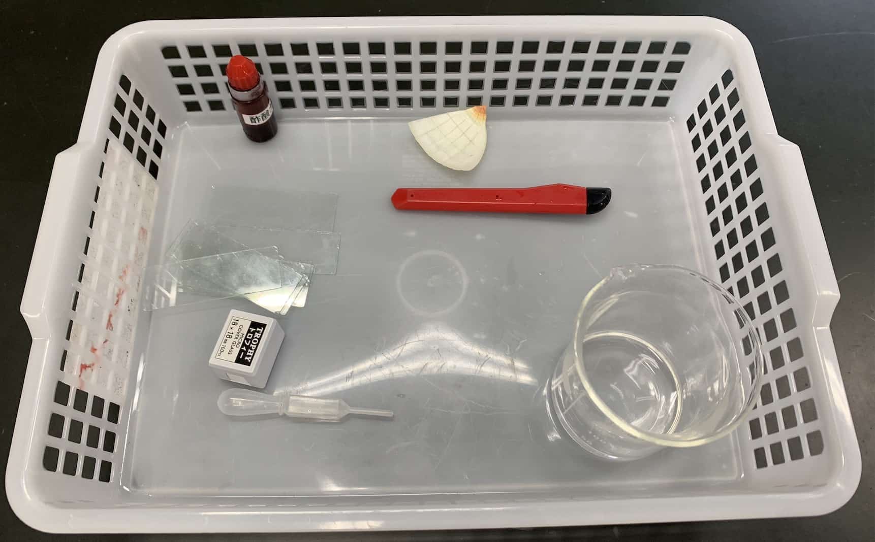
To get started, you will need an onion, two microscope slides, two cover slips, a microscope, tweezers, a staining solution (acetocarmine), a dropper, and a craft knife.
While the kit is simple, the secret to a “eureka” moment lies in the preparation. Most importantly: use a fresh onion. The juicier the onion, the plumper the cells, which makes the view through the lens significantly more beautiful. It’s always thrilling to see an everyday grocery item transform into the ultimate teaching tool.
The “Magic Ritual” of Peeling the Epidermis
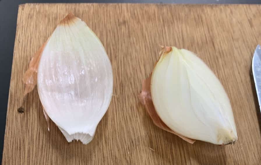
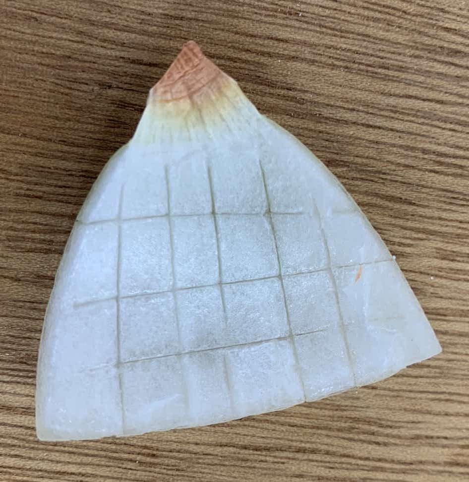
Cut the onion into eighths, peel off a bulb scale, and snap it in half. Then, use the craft knife to make a small square incision (about 5mm) on the inner surface.
This tiny incision is the golden trick to peeling the thin skin (epidermis) smoothly. Letting students adjust the depth of the cut through trial and error is a wonderful first step into the scientific method.
Gently peel away the epidermis along the incision using your tweezers.
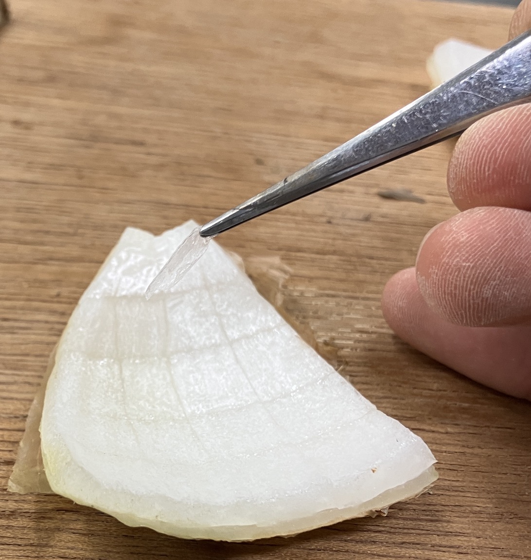
At this point, try asking: “Why does an onion have such a thin skin?” This delicate film is actually a “living barrier” that helps the onion store water underground and protects it from predators and dehydration. Despite being so thin, the way each cell is tightly locked together like bricks is a true masterpiece of natural engineering.
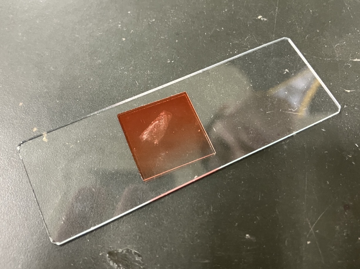
When placing the skin on the slide, the key is to spread it out exactly as it was attached. Avoiding wrinkles ensures the cells don’t overlap, giving you a crystal-clear view of their world. Use a dropper to add a drop of water to one specimen and acetocarmine to the other, then let them sit for about three minutes.
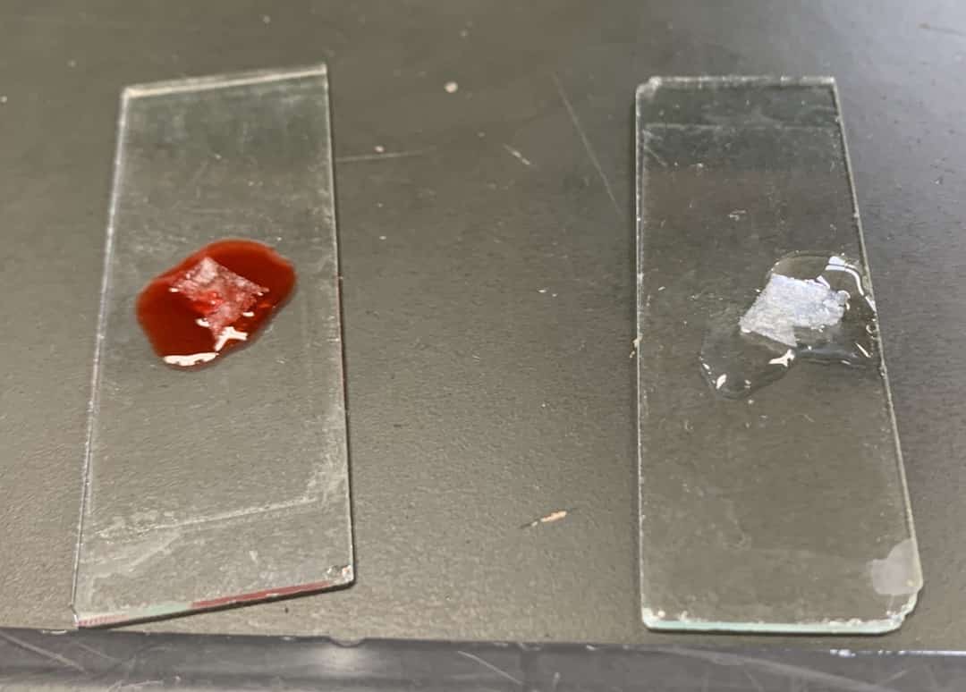
One is “Plain”—to see the onion in its natural state. The other is the “Highlight”—to make specific structures pop. Explaining that this contrast is the foundation of scientific analysis adds a whole new layer of depth to the experiment. Use a mounting needle to carefully place the cover slip, trying your best to avoid air bubbles. If any bubbles get trapped, give it a gentle tap with your tweezers to coax them out. Blot any excess liquid with a tissue.
Peer into the “Cellular Universe”
Now, it’s finally time for the microscope. Start with a low magnification and gradually zoom in. At 400x magnification, students encounter a micro-cosmos that is invisible to the naked eye. You can’t help but feel the incredible order of nature in that perfectly aligned, brick-like pattern.
The Mystery of the Stain: Water vs. Acetocarmine
The highlight of this experiment is comparing the unstained sample with the one dyed by acetocarmine.
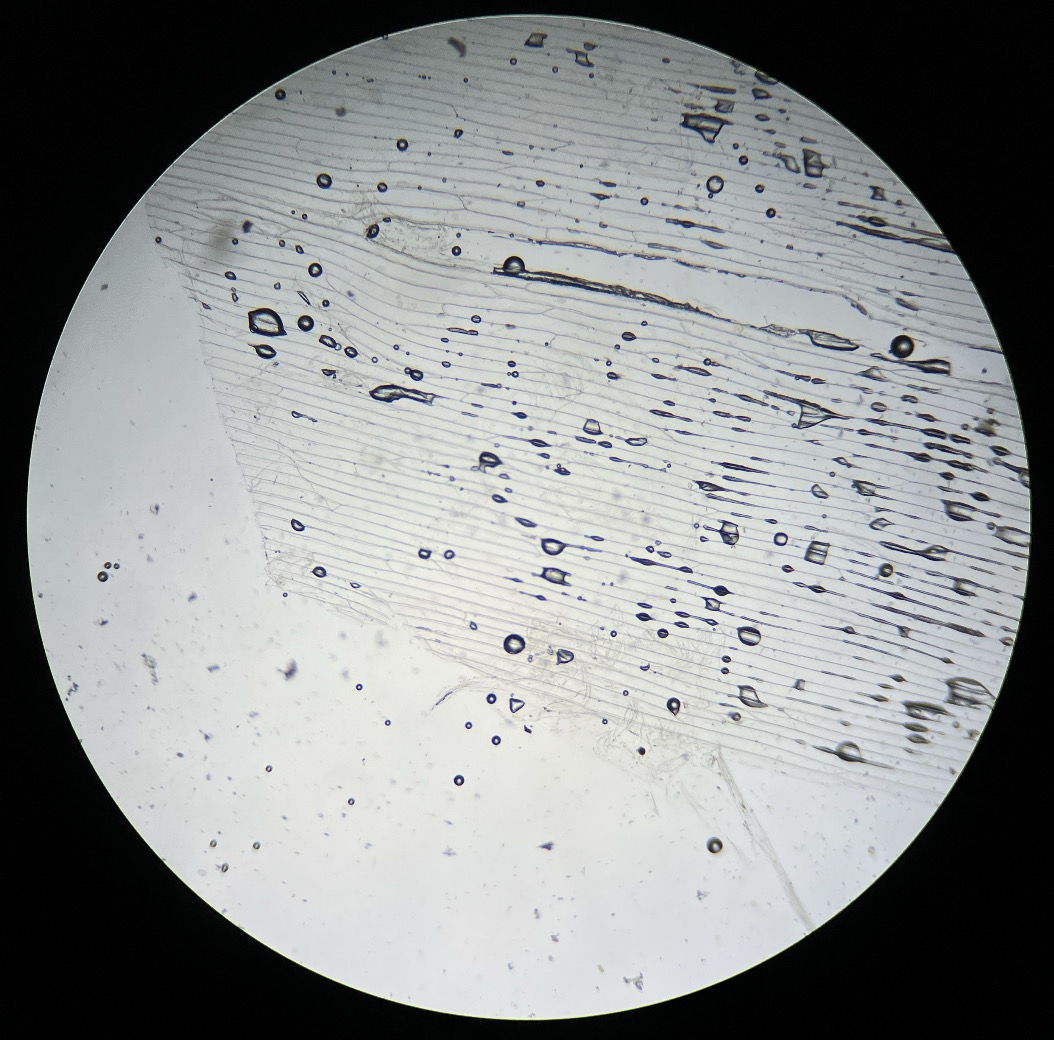
Using Water (Unstained)
(40x: 10x Eyepiece, 4x Objective)
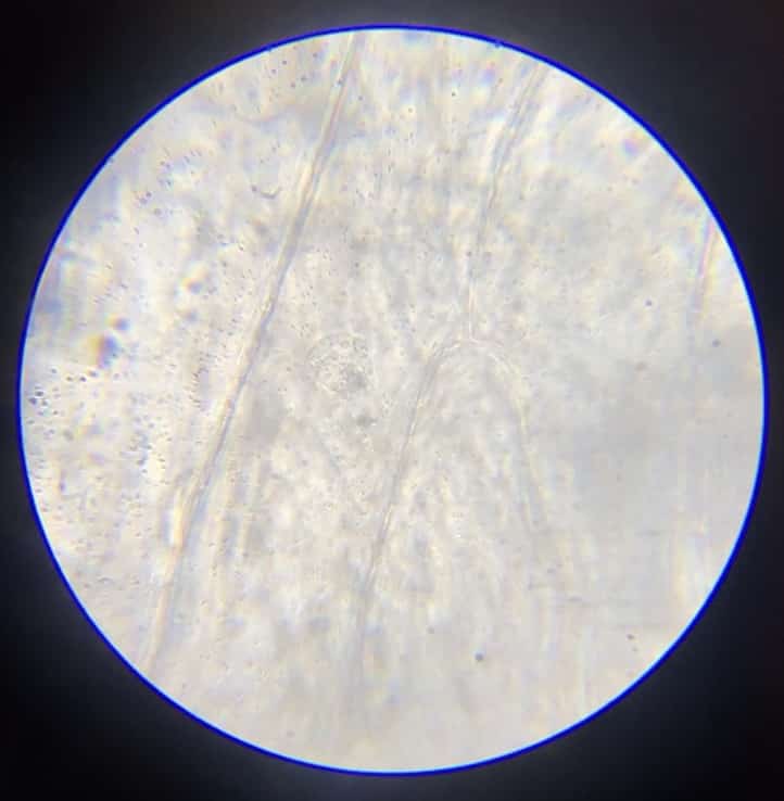
(400x: 10x Eyepiece, 40x Objective)
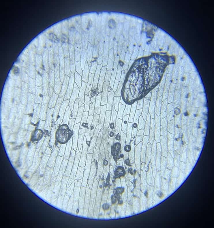
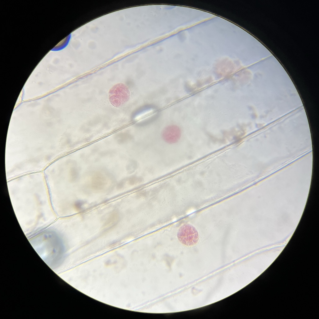
With just water, you can see the outlines of the cell walls, but the vital nucleus remains transparent and blurry. However, when you use acetocarmine, it’s like magic—tiny dots emerge in a vivid red.
Stained with Acetocarmine (100x)
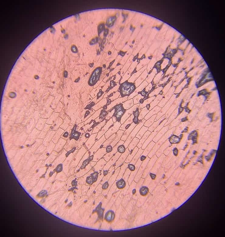
(400x)
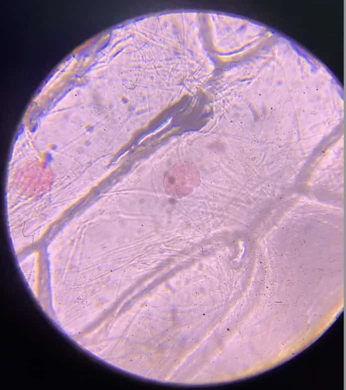
This is the nucleus, the “control center” packed with DNA—the blueprint of life. Here’s a fun fact: why does only the nucleus turn red? It’s because the DNA inside the nucleus carries a negative electrical charge, while the pigment in acetocarmine carries a positive charge. Just like magnets attracting, the pigment is drawn to the DNA, making the nucleus stand out clearly. That moment when the “invisible” suddenly becomes “visible” is the pure joy of science.
Here is another set of results:
40x
100x
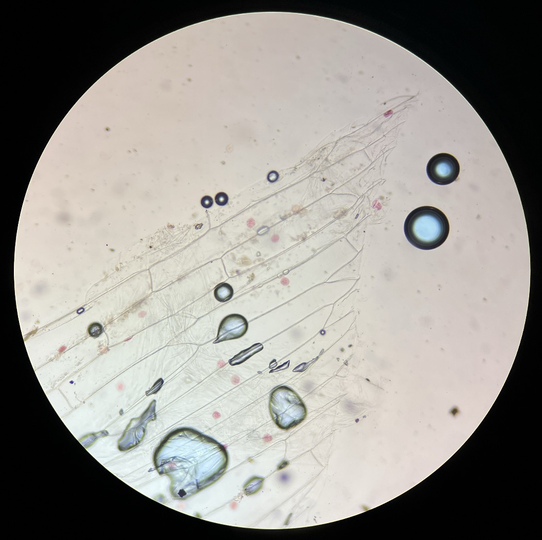
400x
A sophisticated system of life hidden inside a common onion. I hope these small discoveries right in front of you lead to a lifelong passion for science.
Inquiries and Services
Let’s make the wonders of science more accessible! I share tips and fun experiments you can try at home. Feel free to explore my site!
Learn more about the author, Ken Kuwako, here.
For inquiries (writing, speaking engagements, science workshops, TV supervision/appearances), click here.
– Stay updated with my latest posts on X (Twitter)!
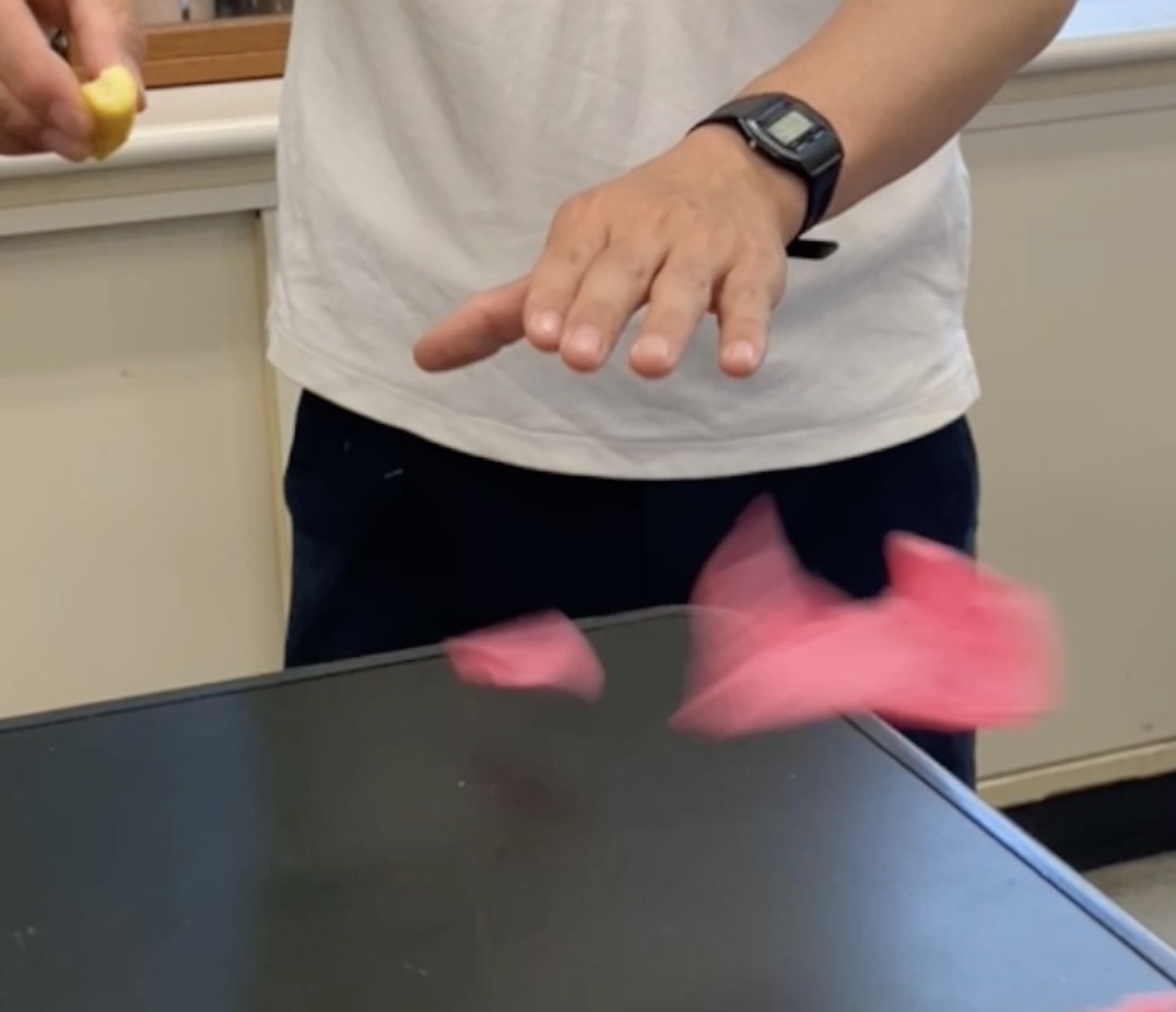
6月のイチオシ実験!
レモンやオレンジで風船を割ろう!インパクトが抜群のリモネン風船の実験
テレビ番組監修・イベント等のお知らせ
- 6月3日(水)20:30〜 「
バカリズムのちょっとバカりハカってみた!」(テレビ東京)を科学監修・出演します。テーマは「 そばの出前は何人前まで運べるのか、限界を測ってみた」です。 - 6月4日(木) 7:00〜 「THE突破ファイル」(日本テレビ)について科学監修しました。
- 6月14日(日) 千葉大学インスタレーション「探究」にて講師を務めます
- 6月26日(金) 公開研究会「脱作業化!デジタル化と段階的指導で実現する オームの法則の探究」
- 6月28日(日) ダビンチマスターズ@昭和女子
- 7月18日(土) 教員向け実験講習会「ナリカカサイエンスアカデミー」の講師をします。お会いしましょう。
書籍のお知らせ
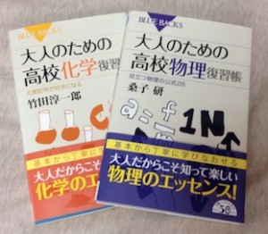
- 『大人のための高校物理復習帳』(講談社)…一般向けに日常の物理について公式を元に紐解きました。特設サイトでは実験を多数紹介しています。※増刷がかかり6刷となりました(2026/02/01)
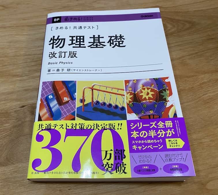
- 『きめる!共通テスト 物理基礎 改訂版』(学研)… 高校物理の参考書です。イラストを多くしてイメージが持てるように描きました。授業についていけない、物理が苦手、そんな生徒におすすめです。特設サイトはこちら。
各種SNS(更新情報をお届け!)
X(Twitter)/instagram/Facebook(日本語)
Explore
- 楽しい実験…お子さんと一緒に夢中になれるイチオシの科学実験を多数紹介しています。また、高校物理の理解を深めるための動画教材も用意しました。
- 理科の教材… 理科教師をバックアップ!授業の質を高め、準備を効率化するための選りすぐりの教材を紹介しています。
- Youtube…科学実験等の動画を配信しています。
- 科学ラジオ …科学トピックをほぼ毎日配信中!AI技術を駆使して作成した「耳で楽しむ科学」をお届けします。
- 講演 …全国各地で実験講習会・サイエンスショー等を行っています。
- About …「科学のネタ帳」のコンセプトや、運営者である桑子研のプロフィール・想いをまとめています。
- お問い合わせ …実験教室のご依頼、執筆・講演の相談、科学監修等はこちらのフォームからお寄せください。