From Humble Moss to Star! Witness Male & Female Liverworts and Their Spring-Loaded Spores Under the Microscope
I’m Ken Kuwako, Science Trainer. Every day is an experiment.
Are You Overlooking the Microscopic Forest at Your Feet? The Fascinating World of Liverworts
Do you ever pay attention to the green ‘moss’ spreading across the roadsides and forgotten corners of schoolyards you walk past every day? Bryophytes—the group that includes mosses and liverworts—are actually like living fossils, carrying the ancient history of when plants first moved from the sea onto land.
When teaching plant reproduction in science class, these non-vascular plants become a profoundly interesting subject for observation. This time, we used the common liverwort (*Marchantia polymorpha*) found on our campus to observe the dramatic structural differences between the male and female plants, and the ‘spores’ that guarantee the next generation. The microscopic world seen through a microscope is guaranteed to amaze your students!
Let’s Collect Liverworts! The Reason They Love Damp Places
Liverworts thrive in moist areas and are often found growing naturally near water features or on shaded, damp ground around the school. Why the preference for wet places? This is because bryophytes haven’t yet developed vascular bundles (the internal ‘plumbing’ for water transport), so they must absorb water directly through their body surfaces. Additionally, as we’ll explain shortly, water is absolutely essential for fertilization.
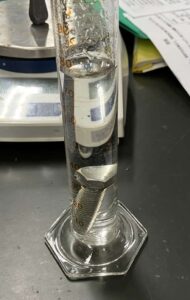
We carefully collected our specimens using a small trowel. When collecting, make sure to take the entire plant, including the root-like structures (rhizoids), and wrap them in a damp paper towel or paper to keep them from drying out.
A Clear Difference! The Distinct Shapes of Male and Female Plants
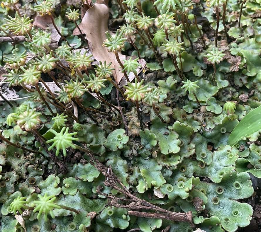
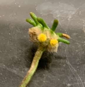
Take a good look at the liverwort you collected. It turns out that, just like humans, liverworts have distinct ‘male’ and ‘female’ plants. And their shapes are surprisingly different from each other.
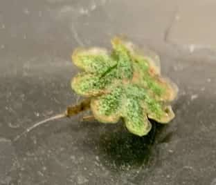
- Male Plant (Antheridiophore): The flat, leaf-like body ends in a flat, disc-like or mushroom-cap-like structure with a scalloped edge. This is where the sperm are produced.

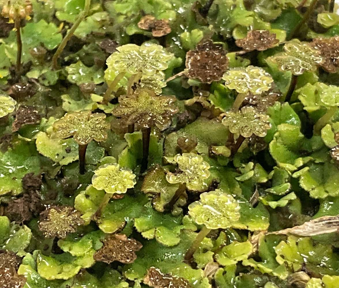
- Female Plant (Archegoniophore): This plant features a protrusion shaped like a palm tree or a tattered umbrella (with finger-like lobes). The egg cells wait underneath this structure, and this is where spores will eventually form.
Their shapes are entirely different, aren’t they? The liverwort thus possesses clear distinguishing characteristics between its male and female forms. But why do they look this way? On a rainy day, when a raindrop hits the male ‘saucer,’ the force of impact disperses the sperm, which then swim through the water to reach the female plant. That flat disc is thought to act as a ‘launch pad,’ utilizing raindrops to propel the sperm! When students observe them while imagining this biological drama, their level of engagement instantly skyrockets.
Spores: The Next Generation of Life, and the Spring-Loaded ‘Elaters’ That Launch Them
Beneath the female ‘umbrella’ is a yellowish, fluffy section that contains the spores. We tried gently tapping this area with tweezers to extract and observe the spores under a microscope. This tiny structure is packed with botanical ingenuity.
Observation Method
Materials Needed
- Mature female liverwort.
- Tweezers.
- Slide glass, cover slip.
- Microscope (400x magnification recommended).
Procedure
- Extract the Spores
- Gently pinch the yellowish, cottony part of the female thallus containing the spores with tweezers, and tap or tease it apart lightly over the slide glass.
-
- Let the spores fall onto the slide glass.
- Prepare the Slide
- Add a drop of water and gently place the cover slip over it.
- Observe under the Microscope
- First, focus using low magnification, then observe the details at 10x eyepiece × 40x objective (400x total magnification).
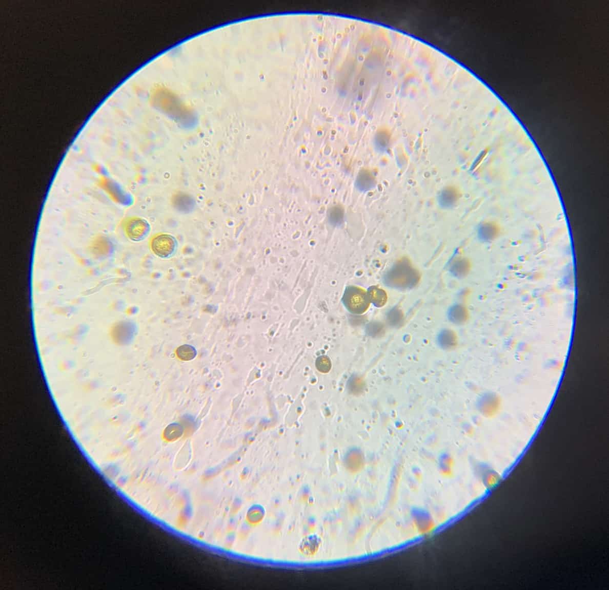
Observation was performed at 10x eyepiece × 40x objective, for a total of 400x magnification.
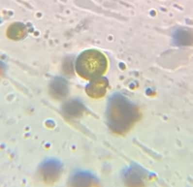
Observation Results: The Identity of the ‘Threads’ Seen with the Spores
Peering into the microscope, you’ll see countless tiny, round granules: these are the spores . And, scattered among the spores—especially if you observe at a higher magnification—you might spot slightly longer, twisted, thread-like structures. These are tissues called elaters . They actually act like a spring , contracting and expanding as the humidity changes. When they dry out, this spring mechanism snaps, launching the spores far away! This is a brilliant physical mechanism non-moving plants have developed to expand their habitat.
Discover the Grandeur in the Tiny Life at Your Feet
Observing the liverwort allows us to explore the vast diversity of plant reproduction and the clever, adaptive mechanisms they use to survive. Mosses and liverworts are often dismissed as ‘gross,’ but when students see their intricate, engineering-like structure under a microscope, their entire perspective changes. This hands-on experience of collecting and magnifying the specimens will surely transform dry textbook knowledge into ‘living knowledge.’
Contact and Requests
Let’s make the wonders and fun of science more accessible! I’ve put together fun science experiments you can do at home and clearly explained the tricks behind them. Feel free to search around! ・About the administrator, Ken Kuwako: Click here ・For various requests (writing, lectures, science workshops, TV supervision/appearances, etc.): Click here ・Article updates are posted on X!
![]() Science Neta Channel is distributing experiment videos!
Science Neta Channel is distributing experiment videos!
5月のイチオシ実験!
キーンと冷えるドライアイス!気温が上がってくるこの時期・ドライアイスを使った昇華・凝結・等速度直線運動の実験はいかが?
液体ゼロ!ドライアイスが消えるまでの3時間を科学する(昇華・凝結・等速度直線運動)
テレビ番組監修・イベント等のお知らせ
- 4月30日(木)「THE突破ファイル」(日本テレビ)の科学監修を担当しました。
- 5月8日(金)理科教育ニュースを担当しました。
- 6月14日(日) 千葉大学インスタレーション「探究」にて講師を務めます
- 6月26日(金) 千葉大学の公開研究会(中学理科について授業公開予定)
- 7月18日(土) 教員向け実験講習会「ナリカカサイエンスアカデミー」の講師をします。お会いしましょう。
書籍のお知らせ
- 『大人のための高校物理復習帳』(講談社)…一般向けに日常の物理について公式を元に紐解きました。特設サイトでは実験を多数紹介しています。※増刷がかかり6刷となりました(2026/02/01)

- 『きめる!共通テスト 物理基礎 改訂版』(学研)… 高校物理の参考書です。イラストを多くしてイメージが持てるように描きました。授業についていけない、物理が苦手、そんな生徒におすすめです。特設サイトはこちら。

各種SNS(更新情報をお届け!)
X(Twitter)/instagram/Facebook(日本語)
Explore
- 楽しい実験…お子さんと一緒に夢中になれるイチオシの科学実験を多数紹介しています。また、高校物理の理解を深めるための動画教材も用意しました。
- 理科の教材… 理科教師をバックアップ!授業の質を高め、準備を効率化するための選りすぐりの教材を紹介しています。
- Youtube…科学実験等の動画を配信しています。
- 科学ラジオ …科学トピックをほぼ毎日配信中!AI技術を駆使して作成した「耳で楽しむ科学」をお届けします。
- 講演 …全国各地で実験講習会・サイエンスショー等を行っています。
- About …「科学のネタ帳」のコンセプトや、運営者である桑子研のプロフィール・想いをまとめています。
- お問い合わせ …実験教室のご依頼、執筆・講演の相談、科学監修等はこちらのフォームからお寄せください。